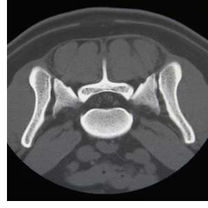
Computed tomography (CT) is a special x-ray technology in which a computer is used to create highly detailed cross sectional images of a portion of the body. In conventional radiography, the x-ray tube remains stationary as an image is generated. In CT, the x-ray tube rotates around the patient and generates axial slices (like the slices in a loaf of bread). CT is especially good in evaluating bone (fractures, infections, tumors) and vertebral lesions such as calcified disks. Neurological computed tomography scans can be used to diagnose fractures, deformities, disease or degenerative conditions. Good quality CT and/or MR imaging is required to completely identify a lesion and its interaction with surrounding tissue (see CT vs MRI). These “blue prints” are especially important prior to surgery to help provide the surgeon with an accurate picture of the problem. During a CT scan, general anesthesia is administered in order to keep patients still while skilled technicians monitor the patient closely during the procedure. A contrast agent (dye) is often administered intravenously to highlight and obtain additional detail of tissue within the area of interest.
►Click here to learn more about Computed Tomography (CT).
This information is meant to be a guide and not a substitute for veterinary care.
Always follow the instructions provided by your veterinarian.
Diagnostic Uses of CT Imaging:
Brain:
- tumors
- hydrocephalus
- inflammation (encephalitis, meningitis)
- vascular lesions (stroke or hemorrhage)
- biopsy (CT-guided)
Spine and Spinal Cord
- tumors
- vascular injuries
- intervertebral disk herniations
- infections
- fractures
- dislocations
- biopsy (CT-guided)
Non-Neurological Imaging
