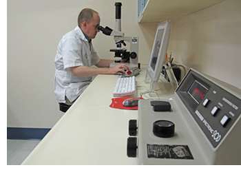
Proper examination of blood, urine, cerebrospinal fluid (CSF) and nervous system tissues is important in evaluating the overall health of a patient as well as providing signs to the cause of certain neurological disorders. For immediate results, several lab tests are performed on-site at the Veterinary Neurological Center. Many off-site laboratories are utilized for less time sensitive results or for tests which may require special equipment.
On-Site Laboratory Testing
Preanesthetic Blood Tests
The Veterinary Neurological Center requests that each patient have a chemistry panel and complete blood count (CBC) performed prior to undergoing any anesthetic procedure. Often this has recently been performed by the referring veterinarian but if it has not or if there is reason for concern, the neurologist at the VNC will run an in-house chemistry and complete blood count (CBC) or, if necessary, send a blood sample to an off-site laboratory for testing. In all cases, however, an in-house preanesthetic panel is always performed prior to anesthesia.