 |
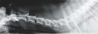 |
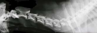
Myelography uses the same technology as radiography, but is specific to the neck and back and utilizes a dye to highlight and outline the shape of the spinal cord. Myelography is used to evaluate a variety of conditions such as herniated disks, spinal tumors, vascular malformations (blood vessel abnormalities) and narrowing of the spinal cord related to degeneration, disease or trauma to the spinal column. General anesthesia is administered to keep patients motionless and technicians closely monitor the patient. A needle is inserted into the subarachnoid space around the spinal cord and cerebrospinal fluid (CSF) flows through the needle. The CSF is collected for possible future CSF analysis. The dye is injected into the subarachnoid space to highlight the spinal cord. The procedure is usually complete within 10 minutes and then spinal radiographs and CT or MR images are obtained.
Myelography is no longer routinely used at the Veterinary Neurological Center because the alternative, MRI, is non-invasive, requires less anesthetic time and provides more detailed views of the spinal cord.
►Click to learn more about myelography.
This information is meant to be a guide and not a substitute for veterinary care.
Always follow the instructions provided by your veterinarian.